Photography & sketching in forensic practice Sharma B. R.1,*, Dr.M.D., Reader, Singh Virendar Pal1, Dr., M.B.B.S., M.D., Demonstrator, Bangar Sumedha1, Dr., M.B.B.S., Demonstrator 1Dept. of Forensic Medicine and Toxicology, Govt. Medical College & Hospital, Chandigarh-160030 *Correspondence: Dr. B. R. Sharma, # 1156 -B, Sector- 32 B, Chandigarh-160030.E-mail: drbrsharma@yahoo.com & drbrsharma@gmail.com
|
INTRODUCTION The use of photographs to determine people's identity has been applied since the middle of the 19th century and included in individual's criminal and prison records1,2-3. With the development and establishment of fingerprint technology, fingerprints became more widely used and proved to be more reliable method of identification. However, even today, throughout the world, photographs are still used and are included as part of prisoner's records. Law agencies universally, routinely use fingerprints and photographs for criminal records. These images commonly referred to as ‘MUG SHOTS’ are characterized by two photographs, one facing directly into the camera and the other a profile. This persistent use of photographs raises the question, as to why photographs are used so commonly when fingerprints are a more reliable source of identification? The answer is quite simple; photographs are used so that a lay person, including police or customs officials, can make cursory identification by comparing the suspect in question with his/her photographs. Fingerprint identification on the other hand, requires a trained and qualified eye viz. a finger print expert4. |
Traditionally, the use of photography in the practice of forensic medicine has been limited to crime scene locations and autopsy suites. But recently, forensic photography is finding its way into emergency departments despite the fact that the status accorded to the scientific evidence in our country is still that of corroborative evidence and not direct evidence, unlike in the developed countries. It has been well observed that, “we still go in for the evidence of an eyewitness, who can be easily manipulated by one and all, thus jeopardizing the case at the outset leading to a very high rate of acquittal by the courts” (State of Karnatka Vs Pundalik, 1999 Cr. L J 4751 Kant). The reasons for employing a camera to aid in addressing the forensic needs include the following: |
To record and document injuries and evidence that cannot be preserved indefinitely or left untouched. To act as a future reference or aid to memory. To document lesser features and details of a situation. To document injuries or conditions, and record what they looked like before and after medical treatment. To show the condition of evidence or injuries at the time of discovery or examination. To demonstrate the absence of alleged injury or finding (s). To illustrate and supplement the written medicolegal and/or medical record. To present in a court of law, the things as they were and, in effect, check the testimony being presented. To teach the undergraduate and postgraduate medical students.
It has been documented that “the evidence of a doctor conducting postmortem examination without producing an authority in support of the opinion is insufficient to grant conviction to an accused” (Mohd. Zahid Vs State of Tamilnadu 1999 Cr. L J 3699 SC). There is sufficient authority for a proposition that “a court is not obliged to accept the opinion of experts even when the evidence has not been challenged by the opposite party in a trial”. Courts have rejected the testimony of experts from Appellant/Defendant on the grounds that the facts on which their opinions were based were not proven. |
Time and again it has been stressed that every forensic expert not only needs to make proper observations but also has to show that it has been done properly. The best way to achieve it is through complete, legible & proper documentation. Proper photographs if taken at the time of a medicolegal autopsy can convey to the court in an appropriate manner what the words fail to convey, although their actual status may have to be decided latter as per the court procedures applicable for evidence. In our legal system, “admission of documents amounts to admission of contents but not its truth” (Afzaudin Ansari Vs State of Bengal 1997). However, a photograph is not usually substantive evidence and will probably be admitted as evidence if: |
It shows the original appearance or findings, and fairly and accurately depicts what the doctor saw. It assists in identification or characterization of the wound or injuries. It is not unduly gruesome or inflammatory. It is of sufficient value to warrant its admission. It will aid the court in obtaining an intelligent or dispassionate evaluation or conclusion. It corresponds closely to the verbal description given by the doctor. It does not contain extraneous matter.
According to National Human Rights Commission's recommendations on Autopsy Protocol, adequate photographs are crucial for thorough documentation of the findings during an autopsy: |
They should be in color, in focus, adequately illuminated and taken by a good quality camera, preferably by a professional. Each photograph should contain a ruled reference scale, an identifying case name or number. Serial photographs reflecting the course of the essential examination prior to and following undressing, washing, cleaning etc. must be included. Identifying facial features should be portrayed with photographs of a full frontal aspect of the face and right and left profiles of the face with hair in normal position and with hair retracted, if necessary, to reveal the ears. Close-up photographs should be supplemented with distant and/or immediate range photographs to permit orientation and identification of the close-up photographs. All photographs should be comprehensive in scope and must confirm the presence of all demonstrable signs of injury or disease commented upon in the autopsy report.
Photographs with an attached scale have an added advantage and present the findings in a still better manner especially the injuries in cases of assault. The following are general guidelines for using scales of size: |
The scale should be sized in proportion to the subject viz. Centimeters for an injury or wound, inches for weapon of offence, feet for offending vehicle, and so forth. The numbers representing centimeters or inches should be large enough to read in the end product. The scale must be in the plane of interest, not behind it (further away from the camera) or in front of it (nearer to the camera). None of the numbers or letters on the scale should be upside down with respect to the subject matter or other numbers that is when you hold the scale in your hand, every thing should be in a single orientation. The placement of the scale should be in a natural orientation, so that it is in a normal reading position when you look at the subject. There should be no advertising material of any kind on the ruler or scale. The scale of size should not compete with the subject matter for attention and should be aesthetically pleasing.
It is worth noting here that if you do not put a scale in a photograph, an attorney might complain, and if you do include one, it might be possible for him to claim that it was covering something important. As such it will be better that the photographs are taken both with and without a scale. |
Conversely, one must also be ready for legal challenges regarding the admissibility of the photographs. The questions and challenges may include: |
Exactly where and when the photograph was taken. Exactly what the photo depicts. The exact anatomical location. The type of camera and film used. If filters were used on the lens. Whether the photographs were obtained by informed or implied consent. Where the film was developed and if it represents the hospital's standard protocol.
The issue of photograph's potentially inflammatory content is likely to be questioned. If a photograph is so gruesome and inflammatory that it may cause ‘undue prejudice’ towards the accused on the part of the court the same may be excluded from evidence. In general, a court is unlikely to admit the photograph if: |
The composition is absurd because the photograph was taken at such a distance from the wound that the wound occupies only a fraction of image. The remainder of image area is taken up with parts of the resuscitation or autopsy table, on which there are a number of potentially offensive and objectionable items, such as bloody sponges, dressings, stains of partly dried blood, scissors etc. The actual wound is out of focus and hard to see. The picture is over-exposed/under-exposed. Poor ‘scale’ technique is used, viz. (a). only a section of plastic ruler is used, cut so that numbers begin at 2 on the inch side and 5 on the centimeter side. (b). An equipment maker's name is on the scale. (c). The scale is covering part of the wound. (d). The scale is not in the plane of the wound but at an angle to it, meaning that since part of the wound is nearer the camera than the scale, any measurement derived from it will be in error by several percentage points. The photograph was not taken at 90 degrees to the wound and perspective distortion is present.
Accordingly, the individuals who intend to take forensic photographs should have some basic working knowledge of photographic theory. Even if they are using a ‘point and shoot’ camera, they will need some background information regarding lens characteristics, aperture, filters, shutter speeds, exposure, depth of field, lighting, background, camera body, film - quality and processing etc. The use of markers may be made to indicate the position of small items in a photograph. However, two photographs, one with and one without a marker should be taken. |
Various areas of medicolegal practice where photographs can be of enormous significance include: |
Scene of Incident: One of the most crucial aspects of the investigation of a suspicious death is the comprehensive examination of the place of the discovery of the dead body. It should be appreciated that the scene at which body is found may not be the same where death occurred. Findings of a study5 are tabulated below in support of this argument. |
An accurate record of the scene is vital for the investigation and subsequent use as evidence in the court. Photographs of crime scene with reference to the body as it is found before and after it has been moved, best serve the purpose. Aerial Photography has been used to photograph out-door scenes of occurrence. It allows better appreciation of the objects at a crime scene. |
Cause of death: It has been emphasized in literature that an autopsy must be complete and meticulous, as the evident cause of death may not always be the real cause of death. The following reports explain the role of photographs in such cases: |
An apparently healthy young man aged 21 years collapsed while jogging in the morning and died before any medical aid could be provided to him.6 Family members suspected homicidal poisoning, while doctor conducting the autopsy suspected Hypertrophic Obstructive Cardiomyopathy. The negative report of chemical analysis and confirmation of HOCM on histopathological examination established the cause of death. However, the photographs of heart, taken at the time of autopsy prove helpful in teaching the undergraduates in such rarely reported cases. A middle-aged person who sustained head injury in 1993, was hit over the abdomen in a scuffle with his neighbor in 1999 (6 years after the head injury). He was shifted to hospital in an unconscious state and died the same day. The detailed autopsy revealed a re-bleed from Chronic Subdural Haematoma as the cause of death7. The photographs attached with the autopsy report rescued the person booked for murder in the court of law. In a case of poisoning with Nitric Acid, the chemical analyzer reported “No poison detected on chemical analysis of viscera”.8 The photograph showing xanthoproteic reaction supported the opinion regarding cause of death.
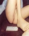
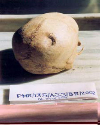
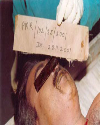
Manner of death:Photograph No.1 showing bruising around the anal opening conveys the findings in a victim of sodomy better than any amount of words9 and Photograph No.2 showing a depressed fracture over the right parietal area of skull of an exhumed body conveys the manner of infliction of injury, type of weapon used as well as the relative position of the assailant and the victim at the time of incident, without using any words. Photograph No. 3 shows xanthproteic reaction along the margins and sides of mouth in a victim of nitric acid poisoning. |
Time since death: Estimation of time of death is a part of the medicolegal inferences drawn after any postmortem examination, though in reality, the investigating police officer is more concerned with the time of assault than the time of death. Estimation of time since death, however, can directly or indirectly help to find out the time of assault. For example, it can help directly, when the death is instantaneous/immediately after the assault, or indirectly, by additional findings related with the stages of healing of an injury. Thus time since death has legal importance in any circumstances. Photographs can provide a record of such changes better than the explanation. |
Postmortem artifacts mean alteration, modification, addition or deletion of some postmortem features, due to certain causes originating after death viz. resuscitation attempt, transportation, preservation in mortuary etc. Postmortem ant bite, cockroach bite and rodent bite injuries may be wrongly interpreted as antemortem injuries by the investigating officers as well as the relatives of the deceased. Photographs in such circumstances can be used to convey/communicate to the court the genuineness of the observations made by the autopsy surgeon. |
Recent Advances Photogrammetry is being used increasingly in some countries to photograph the scene of occurrence. The main advantage of the technique is that it dispenses with the measurement of inter-distances and preparation of sketches. The photograph provides the necessary data on further processing. Digital camera does offer some advantage over traditional slide-and-print films. Digital images are easily stored, e-mailed, and exported into multimedia presentations. The photographer knows within seconds if the image is in focus, well illuminated, well composed and displays what the photographer intended. A digital image can also be easily converted to a slide or print. The digital image can be easily manipulation either for enhancement or alteration. For the last decade, the use of digital enhancement technique has been embraced for improving poor quality and problematic images associated with forensic evidence. In 1990, a digital enhancement process helped resolve a bloody fingerprint on a pillowcover10. Recently, in the developed countries, digital enhancement is being applied to a greater variety of forensic image needs e.g., shoeprints, footprint, tyre-marks, pattern evidence and bite-mark evidence11. Reflective Ultraviolet photography (RUV) is useful to capture the details associated with the surface of the skin and highlights the surface aberrations associated with the injured skin. Since ultraviolet light (UV) penetrates only a few microns into the surface of the skin, capturing photographic images utilizing RUV requires the use of a light source that emits a strong band of UV light, a band-pass filter on the front of the lens that allows only UV light to reach the film, a film that has sensitivity to the UV spectrum and a lens that does not have a filter to block the transmission of UV light. Most commercially available photographic lenses used for visible light photography filter out both UV and infrared (IR) light as these wavelengths are undesirable in visible light photography. Infrared photography can help capture the photographs of injuries below the skin for example sub-cutaneous extravasated blood as the IR light is capable of penetrating up to 3mm below the surface of the skin and it is strongly absorbed in blood. However, the technique requires a special setup as in case with RUV. The focus point for IR photographs is below the surface of the skin and does require a focus shift from the visible focus. Fortunately, many lenses have the IR focus shift marked on the lens and as such the focus correction can be easily accomplished. The IR photographs, when viewed in comparison to UV or Visible light photographs, will appear slightly fuzzy looking because the image being photographed is not on the surface. Sketching: Sketches are handy in depicting a scene of occurrence. In combination with the photographs, the sketches provide an ideal presentation of the scene. They indicate inter-distances between relevant objects. They indicate relevant evidence only.
Top THE FOLLOWING PRINCIPLES SHOULD BE OBSERVED WHILE SKETCHING THE SCENE The sketch of the scene of occurrence should be prepared at the site and not at the office or the residence of the investigating officer. The distance should be measured with tape and not by paces. A suitable scale should be used and indicated on the sketch. The following scales are suitable: For indoor scene: 1:5 to 100; for building and outdoor 1:50 to 500; for large areas - 1:1000 to 10,000. The directions should be indicated. A compass may be used to find out the directions. Superfluous material should not be introduced in the sketch. Symbols, letters or digits and a legend should be used to avoid crowding.
Sketching does not required elaborate equipment. A measuring tape, a drawing boards, a ruler, a set of triangles, graph and drawing paper, pencils and an eraser should ordinarily meet all the requirements. There are various methods of drawing sketches. The following are common: |
Top CO-ORDINATE METHOD The technique is most frequently used. A focal point is chosen and two lines crossing each other at right angle are drawn. One line (x) represents the length and the other (y) the width of the scene to be covered. The location of the objects is then filled by determining the positions of the objects with reference to these co-ordinates. |
Top POLAR TECHNIQUE It is used for large outdoor scenes. Here instead of drawing co-ordinates, the distances and directions of the evidentiary objects are noted with reference to a central (focal) point. The positions are recorded on the sketch accordingly. |
Top SKETCHES IN AUTOPSY REPORTS Sketches can be used to show the shape and location of injury on the body in case the facilities for photography (as is common with the doctors working in rural areas) are not available. They can be handy in explaining the injury to the court but in no case serve the purpose served by photographs. Presently, the sketches are more commonly used to depict the effected and the escaped surface areas of body in cases of patients/victims of burns. |
Top RECONSTRUCTION OF FACIAL CONTOUR Sketching the face of a missing person particularly of an absconding criminal based on the detailed description from persons who have seen the absconding person can help to establish the identity of the missing/absconding person. |
Top CONCLUSION Sketches and photographs can convey in a very simple manner what hundreds of properly coined words may not be able to convey. According to Andreas Feininger, “a good photograph is one which conveys to the observer something which he has not seen, known, or thought of before - - -.” However, when using photographs in forensic practice never purport to be or permit yourself to be qualified as an expert, when in a court of law. One may acknowledge that he has taken some training and/or has knowledge over and above that of a common man. Be qualified as a practitioner, if that is appropriate as opposed to an expert and stay well within your field of expertise just as you would in medicine. |
Top LEGENDS Figures | Photograph No. 1: showing dilatation of and bruising around the anal opening in a victim of sodomy
|  | |
| | Photograph No. 2: Fracture ala signature of right parietal bone in an exhumed body.
|  | |
| | Photograph No. 3: showing yellowish discoloration along the margins and sides of mouth in a victim of nitric acid poisoning
|  | |
|
Table :
| | Scene of Incident | Place of Death | Place of Body Discovery | No. | % | | Same | Same | Same | 471 | 74 | | Different | Same | Same | 123 | 20 | | Same | Same | Different | 36 | 6 |
| |
|