Clinico-pathological alterations in geriatric dogs Pati Soumyaranjan1, Panda S.K.1,*, Acharya A.P.1, Behera M.1 1Department of Veterinary Pathology, College of Veterinary Science and Animal Husbandry, Orissa University of Agriculture and Technology, Bhubaneswar-751003, Odisha, India *Corresponding author: e-mail: drsusen_panda@yahoo.com
Abstract The present study was conducted to evaluate the gross and microscopic changes in the tissues of dogs dying due to geriatric conditions. During the study period, 15 old dogs were taken into consideration as geriatric dogs during clinical presentation and died at different times. History and clinical findings along with haemato-biochemical alterations were studied in all the 15 cases. Systemic postmortem examination of 7 geriatric dogs was conducted and detailed gross lesion in different organ were recorded. Anemia, anemia associated with dirofilariasis, jaundice, hepatitis, nephritis and chronic enteritis were major cause of death during old age. Important gross changes included pale skeletal muscle, enlarged spleen, cardiac dilatation, necrotic liver, nephritic kidney, thickened intestinal wall etc. Significant histopathological findings related to old age were evidenced like glomerular sclerosis, glomerular atrophy in kidney, bile duct hyperplasia and centrilobular necrosis in liver, depletion of the splenic pulp and thickened epidermis of skin at flank region. Top Keywords Canine, Geriatric, Clinical pathology, Histopathology. Top |
INTRODUCTION Geriatrics or old age is a complex biological process which results in changes in the ability to maintain the living homeostasis. It is a well-established fact that geriatrics is naturally selected and genetically programmed in all species of animals1. It is a heterogeneous process which ultimately leads to progressive reduction of ability and gradual onset of morbidity. The most common and first sign of ageing is slowing down2. Old age is manifested with change in behavioral pattern to different visible changes. Hematological and biochemical alterations accompanied by structural and functional changes at tissue or cellular levels are encountered during old age signaling the disease process. Signs of ageing are inevitable in older dogs with all these alterations indicating certain illness or disease. |
Geriatric age chart of different breeds as per Goldston3 is a standard reference protocol for primary screening of geriatric dogs. Most of the times we ignore certain parameters-like external visible changes while determining the old age in dogs. External visible changes include loss of elasticity of skin, rough hair coat, thickening of skin at flank region of hind limb, area around eye and behind the elbow and hock joint, dental wear and tear etc. Along with this different haemato- biochemical alterations also indicate the advent of old age in dogs. Haematological alterations range from anemia to decreased PCV etc. Biochemical changes include decreased serum protein and increased cholesterol etc. in such animals. These haemato-biochemical alterations can directly be correlated with different dysfunctions of heart, liver, kidneys, spleen and musculo-skeletal system etc. Geriatric dogs also encounter certain disease conditions-like arthritis, skin effections fibrosis of kidney and liver, glaucoma, arteriosclerosis, brown pigmentation and haemosiderosis of different internal organs. At times these disease conditions are also associated with tumors. All these changes either at systemic or organ level may lead to loss of functionality in the geriatric animals. The animals also lose their efficacy and agility in the later part of their life. Scanty information is available about age related biomarkers in dogs. Identification of senescent cells through X-gal staining procedure using canine skin tissue collected from dead dogs immediately after death can be correlated with ageing process4. |
All the age-related alterations are not very specific for age determination. It is always difficult to have an accurate age estimation of dog during live and dead conditions. Many times dogs die due to old age complicated with systemic dysfunctions which becomes difficult to establish. There is scanty literature in relation to tissue alterations in geriatric animals whereas geriatric study is extensive in its human counterpart. Therefore, the present study was carried out to detect the aging process through gross and microscopic changes in various organs of geriatric dogs which can be useful for determination of geriatrics. The knowledge of systemic alterations can be helpful for effective management of geriatric dogs at the critical period of their life. |
Top MATERIALS AND METHODS The present study on clinic-pathological changes in geriatric or old dogs was undertaken in the Department of Veterinary Pathology and Teaching Veterinary Clinic Complex (TVCC) spanning a period of about one year. For screening of geriatric dogs, standard geriatric age chart of different breeds as per Goldston3, viz. external visible changes and age as per record were considered as primary screening parameters. During the study period, 15 old dogs were taken into consideration as geriatric dogs during clinical presentation and died at different times (table 1). History and clinical information along with haemato-biochemical alterations were studied in all the 15 cases. Postmortem examination of 7 was conducted and in rest 8 cases, post mortem could not be conducted due to field limitations. Systemic postmortem examination of all 7 dogs was conducted and special care was taken to record the detailed gross lesion in different organs. For histopathological study, representative tissues from different organs with or without gross lesions were collected in 10% formal saline solution. The formalin-fixed tissues were processed by routine histological techniques5. The fixed tissues were washed overnight in running tap water and dehydrated in ascending grades of alcohol and cleared in xylene. |
Paraffin blocks were prepared and sections were cut at 5 micron thickness and stained by haematoxylin and eosin method. Stained slides were examined under microscope for histopathological changes. |
Top RESULTS For screening of geriatric dogs, standard geriatric age chart of different breeds as per Goldston3 was followed. External visible changes and age as per record were considered as primary screening parameters (Table. 1). History and clinical information along with haematobiochemical alterations were studied in all the 15 clinically presented cases (table 2). Clinical findings, hematological and biochemical changes were correlated with the systemic dysfunctions of respective cases associated with old age. Pathomorphology of the respective cases were studied to correlate with systemic dysfunction associated with old age. All the seven cases were grouped into various suspected systemic dysfunctions on the basis of clinical and haemato- biochemical findings as: (1) anemia -2, (2) anemia and dirofilariasis -1, (3) jaundice, hepatitis & nephritis -2, (4) hepatitis and nephritis -1, and (5) chronic enteritis -1. |
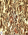
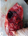
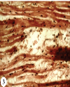
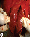
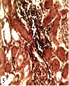
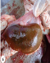
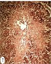
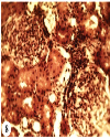
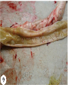
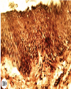
Pathomorphology in anemia Grossly, in both the cases, skin was thickened at flank region of the hind limb, area around eye, behind elbow and hock joint. However, skin was thinner in other regions as usual in geriatric dogs. Oral mucosa, conjunctiva and skeletal muscles were pale. Lungs were pale and edematous. Round heart with right ventricular hypertrophy was also noticed. Spleen was markedly enlarged. Microscopically, epidermis of the skin of the flank region of the hind limb was thickened with one to two layers having few prickle cells. Heart showed congestion of blood vessels and perivascular oedema. In lungs, perivascular and peribronchial spaces were oedematous and parts of the pulmonary parenchyma revealed emphysema. Empty venous sinuses were found in spleen with split in the white pulp area. There was also depletion of Malpighian corpuscles (Fig. 1). Pathomorphology in anemia and dirofilariasis The omentum and peritoneum were pale. Grossly, there was cardiac dilatation with presence of few dirofilarial worms in both the ventricles (Fig. 2). Spleen was enlarged. Microscopically, heart showed oedema, congestion and disruption of muscles in myocardium (Fig. 3). Blood vessels in myocardium showed increased number of leucocyte cells with congestion of vasa vassorum. The trabecular blood vessels of the spleen showed increased leucocytic infiltration. Pathomorphology in jaundice, hepatitis and nephritis Fecal content was yellowish. Grossly, in both the cases, there was yellowish tinged skeletal muscles and subcutaneous fat. Cut surfaces of the liver showed yellowish discoloration (Fig. 4). Stomach was empty with scanty yellowish mucus. Heart was round in shape in one case. Kidneys were pale with slight rough surface and in other one there was marked rough surface. Yellowish tinged perirenal fat was evident in one case. Microscopically, there was focal interstitial infiltration of inflammatory cells in kidney (Fig. 5) and there was hyalinization of tubular epithelium. Liver showed extensive centrilobular necrosis and markedly distended and congested sinusoidal spaces. Hepatic portal region showed bile duct hyperplasia with duplication and thickening of some of the ducts. Pathomorphology in hepatitis and nephritis Grossly, liver was pale with necrotic patches. Kidneys were atrophic with shrunken, rough cortical surface (Fig. 6) and thickened capsule, sometimes showing adhesions with subcapsular surface at places. Microscopically, liver showed extensive centrilobular necrosis (Fig. 7) and bile duct hyperplasia with duplication and thickening of some of the ducts. Kidney was atrophied with thickening of Bowman‘s capsule. There was tubular dilatation with lumen occasionally containing fibrinous exudate, but were frequently empty; tubular epithelium showed degenerative and necrotic changes and pyknosis of nuclei. In cortical region, glomeruli showed swelling and adhesion to Bowman‘s capsule and glomerular sclerosis (Fig. 8). Pathomorphology in chronic enteritis old age Grossly, intestine revealed thickening of the wall containing firmly adhered thick brownish diphtheritic exudate (Fig. 9). At places there was ulceration of mucosal surface, congestion and haemorrhages. Muscles were dry. Microscopically, there was desquamation of epithelial cells of the mucosa at places with presence of necrotic debris. Lamina propria was infiltrated by mixed inflammatory cells. Skin of the flank region showed thickening of epidermis particularly the stratum spinosum layer (Fig. 10). Top DISCUSSION Most organs were altered morphologically and functionally in old animals6. Physiological changes that occur throughout the ageing process reflect the gradual loss of functional activities in various systems7 organ. Grossly, skin was thickened at flank region of the hind limb, area around eye, behind elbow and hock joint. However, skin was thinner in other region in geriatric dogs. The pathological changes observed in various organs in present study on geriatric dogs revealed that heart, liver and kidney were found to be the major affected organ during postmortem which are mostly the metabolically demanded organs8. One of the common symptoms of liver affection is jaundice, a yellowish tinge to the skin, most often noticed in eyes, gums and even on cut surfaces of internal organs which lead to yellowish appearance. Jaundice and icterus are hallmark of hepatic disorder in aged dogs9. Important histopathological findings in kidney were glomerular sclerosis, glomerular atrophy and tubular atrophy. In age related changes of renal histomorphology, tubular atrophy is the common and pronounced findings in aged dogs between 7–14 years old10. It may also include glomerular lesions-like glomerular sclerosis which is in accordance with the above study. Age related changes are quite evident in kidneys that include decrease in number of glomeruli, decreased tubule size and increased renal fibrosis11. The renal lesions mostly the degenerative and inflammatory changes were associated with old age12. There is also reports of renal amyloidosis and immune complex deposition in the glomerulus which is not observed in the present study12. Above kidney changes can be correlated with ageing process. |
In all cases the splenic pulp showed severe depletion of lymphoid cells from the Malpighian corpuscles which indicate decreased immune response that further can be correlated with aging. Aging has been associated with decreased function of the immune system in a variety of species13. Grossly, spleen was also found to be enlarged which might be immune mediated response. The most evident age related effects in the heart were loss of muscles fibers due to disruption of muscle fibres along with congestion of vasa vassorum14. Blood vessels were markedly congested in the intestinal sub mucosa in geriatric dogs14. Geriatric stage is an important aspect of life of a dog during which it encounters different patho-morphological alterations affecting functionality of different vital organs. These pathological alterations must be identitial and addressed in geriatric dogs to augment its quality of living and to enhance the life expectancy of the animal. |
Top Figures | Fig. 1.: Spleen: Depletion of Malpighian corpuscle. H&E ×400
|  | |
| | Fig. 2.: Heart: Presence of dirofilarialworms in the ventricle
|  | |
| | Fig. 3.: Heart: showing oedema, congestion and disruption of cardiac muscle fibres. H&E ×400
|  | |
| | Fig. 4.: Liver: Pale and yellowish cut surface.
|  | |
| | Fig. 5.: Kidney: Focal in infiltration of inflammatory cells interstitial. H&E ×400
|  | |
| | Fig. 6.: Kidney- Nephritis with rough cortical surface
|  | |
| | Fig. 7.: Liver-Extensive centrilobular necrosis. H&E ×100
|  | |
| | Fig. 8.: Kidney: Glomerural sclerosis with swelling of the glomeruli and adhesion to Bowman‘s capsule.H&E ×400
|  | |
| | Fig. 9.: Intestine: Thickened wall with scanty exudates
|  | |
| | Fig. 10.: Skin of flank region: Thickened epidermis at the stratum spinosum layer. H&E ×400.
|  | |
|
Tables | Table 1.: External visible changes and age considered as primary screening parameters
| | Case No. | Breed | Sex | Age (Years) | Visible changes related to old age | Presented for postmortem | | 1 | Labrador | Male | 9 | Thickening of foot pad, rough hair coat and dental wear and tear | Died in one month | | 2 | Golden Retriver | Male | 10.2 | Rough hair coat, Dental wear and tear, thickening of foot pad and halitosis | Died in 21 days | | 3 | Labrador | Female | 10 | Rough hair coat, alopecia and dental wear and tear | Died in 25 days | | 4 | Spite | Male | 8 | Thickening of foot pad, dental erosion and alopecia | Died in two months | | 5 | Doberman | Male | 11.5 | Alopecia, halitosis, dental erosion and wrinkling of skin | Died in 1 month and 10 days | | 6 | German Spitz | Female | 13 | Hair fall, dental wear and tear, halitosis | Died in two months | | 7 | Pomerian | Male | 14.5 | Dental erosion, glaucoma and halitosis | Died in 29 days | | 8 | Spite | Male | 13 | Kearatoconjunctivitis, alopecia, wrinkling of skin and broken teeth | Died in 1 month | | 9 | Spite | Male | 10 | Broken teeth, keratinization of foot pad and alopecia | Died in 1 month and 10 days | | 10 | Labrador | Female | 14 | Glaucoma, dental erosion, yellowing of teeth and halitosis | Died in 21 days | | 11 | pug | Female | 13.5 | Alopecia, dental erosion, yellowing of teeth and glaucoma | Died in two months | | 12 | Pomerian | Male | 11 | Calluses on elbow and knee and graying of hairs | Died in 27 days | | 13 | Labrador | Male | 10.2 | Cataract, poor hair coat and callus formation on knee | Died in 1 month and 12 days | | 14 | Golden Retriever | Male | 13 | Rough hair coat, alopecia and wrinkling of skin | Died in 1 month | | 15 | Labrador | Male | 11.8 | Dental erosion, broken teeth, halitosis and glaucoma | Died in 29 days |
| | | Table 2.: History and clinical findings along with haemato-biochemical alterations in dogs.
| | Case No. | Clinical Findings | Haematological Alterations | Biochemical Alterations | Interpretations | Systemic Dysfunction | | 1 | Pale oral mucosa, conjunctiva and muzzle | Hb (7.1g), PCV (21) TLC (3900) & TEC (3.6) | SGPT (54.3) & SGOT (35.23) | Lower Hb, PCV, TEC & TLC along with rise in SGOT and SGPT values | Anemia | | 2 | Animal was offfed, weak, emaciated and yellowish discoloration of body | Hb (9.1g), PCV(25), TLC (3500) & TEC (3.3) | SGPT (50.38), SGOT (51.06), Creatinine (2.19) & Albumin (1.76) | Lower TLC value along with high rise in SGPT, Creatinine but lower serum Albumin | Jaundice, and Nephritis | | 3 | Pale oral mucosa, conjunctiva and animal was off-fed | Wet B/S- (Positive for microfilaria) Hb (5.9g), PCV (25), TLC (4500) & TEC (3.8) | SGPT (44.33), SGOT (54.33, Urea (45.93) & Creatinine (1.4) | Lower Hb, PCV and TEC along with rise in SGOT and urea | Anemia and Dirofilariasis | | 4 | Animal was off-fed, weak emaciated and yellowish discoloration of mucosa | Hb (9.2g), PCV (26), TLC (3500) TEC (3.5) | SGPT (53.38), SGOT (53.07), Creatinine (2.19) & Albumin (1.67) but lower serum Albumin | Lower TLC along with high rise in SGPT, SGOT and Creatinine | Hepatitis and Nephritis | | 5 | Animal was off-fed, pale skin, dizziness, pale oral mucosa, conjunctiva and shortness of breath | Hb (6.0), PCV (20), TLC (3700) TEC (3.4) | SGPT (52.4), SGOT (33.15) | Lower Hb, PCV, TLC along with rise in SGOT and SGPT values | Anemia | | 6 | Inappetance, animal was weak, decreased painful and urination | Hb (5.9), PCV (20), TLC (3800) & TEC (4.2) | SGPT (53.95), SGOT (51.92) Creatinine (2.22) & Albumin (1.73) | Lower Hb, PCV along with high rise in SGPT, SGOT, Creatinine but lower Albumin | Nephritis and Hepatitis | | 7 | Joint pain, yellowness of the skin, scanty urination, fatigue and nausea | Hb (6.0), PCV (21), TLC (3500) & TEC (3.9) | SGPT (47.95), SGOT (50.92) Creatinine (2.12) & Albumin (1.33) | Lower Hb, PCV along with high rise in SGPT, SGOT, Creatinine but lower Albumin | Nephritis and Hepatitis | | 8 | Vomition, dizziness and animal was weak | Hb (7.4), PCV (26), TLC (29000) & TEC (3.9) | Glucose (73.81), Total Protein (2.95) & Urea (44.19) | Lower Glucose and Total Protein but increase in serum urea value | Enteritis | | 9 | Inappetance, vomition and animal was weak | Hb (8.5), PCV (27), TLC (27000) & TEC (4.9) | Glucose (73.81) Total Protein (2.95) & Urea (44.19) | Lower Glucose and Total Protein but increase in serum urea | Chronic Enteritis | | 10 | Animal was weak and emaciated, off-fed and yellowish discoloration of mucosa | Hb (9.3), PCV (24), TLC (3900) & TEC (3.1) | SGPT (52.38), SGOT (53.77) Creatinine (2.08) & Albumin (1.63) | Lower TLC along with high rise in SGPT, SGOT, Creatinine but lower serum Albumin | Jaundice, Hepatitis and Nephritis | | 11 | Scanty urination, fatigue, loss of appetite, nausea, joint pain and yellowness of the skin | Hb (6.0), PCV(21), TLC (3800) & TEC (4.1) | SGPT (49.95), SGOT (52.92) Creatinine (2.02) & Albumin (1.43) | Lower Hb, PCV along with high rise in SGPT, SGOT, Creatinine but lower Albumin | Nephritis and Hepatitis | | 12 | Animal was weak, inappetance and vomition | Hb (8.7), PCV(26), TLC (29000) & TEC (5.1) | Glucose (71.81), Total Protein (2.92) & Urea (43.17) | Lower Glucose and Total Protein but increase in serum urea | Chronic Enteritis | | 13 | Cloudy urine, loin pain, reduced urine and inappetance | Hb (5.0), PCV (23), TLC (3700) & TEC (4.1) | SGPT (53.85), SGOT (50.93) Creatinine (2.32) Albumin (1.43) | Lower Hb, PCV along with high rise in SGPT, SGOT, Creatinine but lower Albumin | Nephritis and Hepatitis | | 14 | Pale oral mucosa, conjunctiva, animal was off-fed, pale skin, dizziness and shortness of breath | Hb (7.0), PCV(23), TLC (3700) & TEC (3.5) | SGPT (53.3I) & SGOT (34.13) | Lower Hb, PCV, TLC along with rise in SGOT and SGPT | Anemia | | | 15 | Animal was off-fed, pale oral mucosa, conjunctiva and unable to move | Wet B/S-(Positive for microfilaria), Hb (5.8) PCV (25) & TLC (4400) TEC (2.8) | SGPT (44.21), SGOT (51.33) Urea (41.93) & Creatinine (1.3) | Lower Hb, PCV and TEC along with rise in SGOT and urea | Anemia and Dirofi lariasis |
|
| N.B.: Units - Hb g%, PCVC%, TEC×106mm3, TLC×103/mm2, Glucose mg/dl, Total Protein mg/dl, Albumin mg/dl, SGPT IU/L, SGOT IU/L, Cre atinine mg/dl & Urea mg/dl | |
|