Infectious coryza in birds complicated by other bacterial infections Dwivedi Sandeep1, Swamy Madhu1,*, Dubey Amita1, Verma Yamini1, Singh A.P.1 1Department of Veterinary Pathology, College of Veterinary Science and A.H., Nanaji Deshmukh Veterinary Science University, Jabalpur-482001, Madhya Pradesh, India *Corresponding author: e-mail: vetpath@rediffmail.com
Abstract Infectious Coryza (IC), caused by Avibacterium paragallinarum, is an infectious and highly contagious bacterial respiratory disease of poultry. The present communique deals with the pathological investigation of complicated cases of Infectious Coryza. The dead birds with nasal/lacrimal discharge and swollen sinuses were screened for the presence of Avibacterium paragallinarum and other concurrent bacterial infection by PCR and microbial culture, respectively. The birds (n=08) found positive for Avibacterium paragallinarum were also found to be infected with other bacteria like E. coli, Klebsiella, Salmonella and Pseudomonas. Pathologically, fibrinous deposits were seen in airsac, liver and heart of all the birds but more severe changes were seen in birds with concurrent E. coli infection. The histopathological lesions like tracheitis and deciliaton were consistently observed in the trachea, and lungs from all birds with complicated coryza and also showed interstitial pneumonic changes. All these cases were diagnosed as complicated cases of infectious coryza. Top Keywords Avibacterium paragallinarum, Infectious Coryza, Poultry. Top |
INTRODUCTION Infectious Coryza is caused by a Gram negative, non-motile bacterium, Avibacterium paragallinarum.IC is emerging as one of the major problems affecting commercial poultry industry now a days. IC is one of the major problems seen in different part of the country causing significant loss to commercial poultry industry and is found to affect both growing broiler and layer type chickens. The economic impact of the disease is attributed to the resultant increase in unthrifty chickens, significant drop in egg production (10 to 40%) in layer flocks and retardation of growth due to decreased feed and water consumption in breeder and broiler flocks, leading to increased number of culls particularly on multi-aged farms1. When Infectious Coryza is associated with other viral or bacterial infectious agents such as infectious bronchitis virus, Mycoplasma gallisepticum, Escherichia coli, Salmonella spp. or Pasteurella multocida, the disease worsens and prolongs its course and is termed as “Complicated Infectious Coryza”2. Severe and prolonged disease may develop in complicated cases with the clinical picture of a chronic respiratory disease, swollen head like syndrome, airsacculitis, tarsal arthritis and septicaemia. In India, outbreaks of Infectious Coryza are often complicated with fowl cholera and can result in high mortalities (50%) in layer flock3. |
Results of previous studies on outbreaks of Complicated Infectious Coryza have shown the necessity to carry out detailed bacteriological and pathological studies of such cases in different geographical areas2. Study of epidemiology of a disease is important in order to prevent a disease entering a new region or farm, to reduce economic impact produced by the disease and for planning, monitoring and assessment of disease control programmes. Till now, no systematic epidemiological study has been carried out about infectious coryza in Madhya Pradesh, also to our knowledge little research on the pathology of Complicated Infectious Coryza has been conducted in our country. Hence, the present work was taken up to determine the prevalence of avian Infectious Coryza and to study the pathology of multi-systemic lesions in the birds infected with other bacteria along with Avibacterium paragallinarum. |
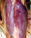
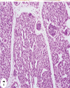
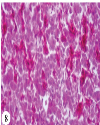
Top MATERIALS AND METHODS Collection of samples Sixty pooled ocular, nasal and tracheal swabs were collected each for molecular and microbiological examination of the dead birds from 20 different broiler flocks showing typical lesions-like oculo-nasal discharge and swollen face suggestive for Infectious Coryza. Swabs for the molecular diagnosis were collected in PBS and stored at 4°C, whereas swabs for microbial diagnosis were collected in Ames transport medium with charcoal. Tissues from various organs-like trachea, lung, liver, heart and kidney were collected for histopathological examination. Molecular detection PCR screening of the samples was carried out using N1(5' TGA GGG TAG TCT TGC ACG CGA AT 3') and R1(5' CAA GGT ATC GAT CGT CTC TCT ACT 3') HPG-2 gene specific primer pairs to amplify 500 bp fragment of Avibacterium paragallinarum4. Reference strains The standard reference strains of Avibacterium paragallinarum (Spross, Modesto and 0083) were procured from Stallen South Asia Private Limited, Mumbai, Maharashtra to use as positive control in PCR. Isolation and identification of other bacteria Swabs from the Ames transport media were revived in the nutrient broth and BHI broth and were further streaked on Nutrient agar. Single colonies from the Nutrient broth were re-streaked on EMB, XLD, MacConkey and Blood agar plates for enrichment. Identification of the bacteria was based on the colony morphology, Gram‘s staining and Biochemical characterization. Gross and histopathology Detailed PM examination was carried out and gross lesions in multiple organs were recorded and pieces of the tissues were collected in 10% formalin. The tissues from the carcasses found positive for Avibacterium paragallinarum were further processed for microscopic study. Top RESULTS Molecular detection of Avibacterium paragallinarum All the 60 pooled ocular, nasal and tracheal swabs were tested along with the positive control for the presence of Avibacterium paragallinarum (AP) using specific primers. Eight samples out of 60 (13.33%) were found positive for Avibacterium paragallinarum and revealed an amplified product of 500 bp (Fig. 1). Isolation of other bacteria from cases positive for Avibacterium paragallinarum The eight dead birds diagnosed positive for Avibacterium paragallinarum were further examined for the presence of other bacterial infections to study their complication in IC. The bacteria were identified on the basis of morphology and biochemical characteristics. Three birds were found positive for mixed Avibacterium paragallinarum, E. coli and Klebsiella spp., two birds tested were positive for Avibacterium paragallinarum and E.coli. two birds for Avibacterium paragallinarum, Klebsiella spp. and Salmonella spp.; and one bird was found positive for Avibacterium paragallinarum and Pseudomonas spp. Gross pathology Gross pathological lesions were predominantly found in facial region and organs of respiratory system in all the cases of complicated Infectious Coryza. The facial lesions observed were conjunctivitis, serous ocular discharge, serous to yellowish brown nasal discharge (Fig. 2), swollen sticky eyelids, swollen face and sinuses (Fig. 3) along with misshapen heads and unilateral or bilateral swelling of wattles. The nasal turbinates were hyperemic filled with thick mucoid and fibrinous exudates. In all the eight cases, congestion and mucoid exudates were observed in tracheal lumen. One bird found positive for both AP and Pseudomonas had caseous plug in the tracheal opening (Fig. 4). The lungs of all the birds were congested and slightly enlarged. Examination of the carcasses for extra respiratory lesions revealed mild to moderate deposition of fibrinous material over heart, liver (Fig. 5) and airsacs. Birds found positive for AP, Klebsiella and Salmonella had enlarged, discolored friable liver, distended abdomen and hemorrhagic enteritis. Swollen hemorrhagic kidney was observed in two birds with AP, E. coli and Klebsiella spp. Histopathology Microscopic examination of mucosal epithelial and glandular cells of the middle and rostral nasal sinuses revealed thickened lamina propria, epithelial degeneration, and hypertrophy of mucosal glands with infiltration of mononuclear cells. Hypertrophy and hyperactivation of mucosal glands were observed in the trachea (Fig. 6). Tracheal lumen showed desquamated epithelial cells. Lung showed interstitial pneumonic changes with congestion, thickened inter bronchial space, focal hemorrhage haemosiderosis and accumulation of fibrinous exudates in the parabronchial lumen (Fig. 7). The section of lung from carcass of birds with AP and E.coli. presented discontinued respiratory epithelium, absence of cilia and hyperplasia of non-ciliated epithelial layer in the secondary and parabronchi. In addition to the respiratory lesions, the sections from heart, liver and kidney were also examined for microscopic pathology. Sections from the heart of the carcasses from birds with complicated IC revealed disrupted muscle fibers, patches of hemorrhages and multiple foci of infiltration of mononuclear cells. Liver revealed bile duct proliferation and cellular infiltration in the portal triad and mild degeneration of hepatocytes along with multiple foci of hemorrhages (Fig. 8). Sections of kidney from maximum birds revealed renal tubular degeneration, glomerulopathy and cellular infiltration. The extra respiratory lesions in liver and heart were more severe in the birds having combined E.coli and AP infection. Top DISCUSSION The present study evaluated the gross and microscopic pathology of various organs of birds with complicated Infectious Coryza. Association of infections with pathological analysis is essential to establish the relationship between cause and effect and to allow the definitive diagnosis5. All the cases found positive for AP were also having concurrent bacterial infections; similar findings were also reported by earlier workers6, so denominated ascomplicated cases of Infectious Coryza. |
Almost similar gross respiratory lesions were observed in all the birds and were similar to those described earlier9,10. No appreciable differences in the severity of respiratory lesions were observed in birds with different bacterial infections. Although the fibrinous deposits were seen in airsac, liver and heart of all the birds but more severe changes were seen in birds with concurrent E. coli infection. E. coli considered as the normal microfloral inhabitant of many avian species11 can also cause different diseases in poultry including respiratory infections12. Klebsiella and E. coli are also reported to induce pneumonia and tracheitis13. Klebsiella being a microbe of low pathogenicity affects the immunosupressed birds in by intensifying the course of primary infections14. |
The histopathological lesions-like tracheitis and deciliaton were consistently observed in the trachea, similar lesions were also reported by earlier workers1,7,11,15. Moreover, dedliation was not common in all the sections as intact cilia were also observed. Increase in number of goblet cells and the size of mucous glands are associated with excess mucous production and may have resulted due to catarrhal inflammation and are reported in the initial cases of respiratory diseases16. Lung sections of all the bird with Complicated Avian Infectious Coryza presented interstitial pneumonic changes supporting the observations of previous workers1,7. |
Avibacterium paragallinarum is known for its pathogenic synergism with other respiratory pathogens including bacteria and viruses. Also the increased severity of the disease was previously reported with co infection of Mycoplasma gallisepticum and E. coli17,18. Infectious coryza primarily affects upper respiratory tract but recently it has been stated that it is not only limited to respiratory tract but it sometimes take systemic form and causes diarrhea8. |
To conclude, the lesions were observed due to the mixed pathogenicity of A. paragallinarum and other bacterial infection. Studies regarding molecular detection, culture and extensive epidemiological studies for allied respiratory pathogens are suggested to know the magnitude of complicated IC infection in Indian poultry flocks and to develop preventive measures. |
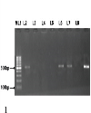
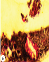
Top Figures | Fig. 1.: Gel electrophoresis showing amplified product of 500 bp of 16s rRNA for identification of Avibacterium paragallinarum in birds with coryza. M: 100 bp ladder, L1, L5 and L6: positive control, L7: negative control, L8: positive control
|  | |
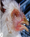
| | Fig. 2.: Layer grower with serous ocular and yellow nasal discharge
|  | |
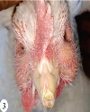
| | Fig. 3.: Bird with severely swollen infraorbital sinuses and wattles suggestive of Infectious Coryza
|  | |
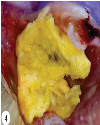
| | Fig. 4.: Caseousplug in the tracheal lumen of IC affected bird.
|  | |
| | Fig. 5.: Bird with Complicated Infectious Coryza having fibrinous perihepatitis and pericarditis
|  | |
| | Fig. 6.: Trachea of bird with Complicated Infectious Coryza having hypertrophy of mucosal glands. Mucicarmine x400
|  | |
| | Fig. 7.: Lung of bird with Complicated Infectious Coryza having congested arteriole and thickened interbronchial septa. H&E ×400
|  | |
| | Fig. 8.: Liver from bird with Complicated Infectious Coryza showing hemorrhages. H&E ×400.
|  | |
|
|